Tissue-Specific Sparse Deconvolution for Low-Dose CT Perfusion
People
Ruogu Fang, Tsuhan Chen, Pina Sanelli
Abstract
Sparse perfusion deconvolution has been recently proposed to effectively improve the image quality and diagnostic accuracy of low-dose perfusion CT by extracting the complementary information from the high-dose perfusion maps to restore the low-dose using a joint spatio-temporal model. However the low-contrast tissue classes where infarct core and ischemic penumbra usually occur in cerebral perfusion CT tend to be over-smoothed, leading to loss of essential biomarkers. In this paper, we extend this line of work by introducing tissue-specific sparse deconvolution to preserve the subtle perfusion information in the low-contrast tissue classes by learning tissue-specific dictionaries for each tissue class, and restore the low-dose perfusion maps by joining the tissue segments reconstructed from the corresponding dictionaries. Extensive validation on clinical datasets of patients with cerebrovascular disease demonstrates the superior performance of our proposed method with the advantage of better differentiation between abnormal and normal tissue in these patients.
Algorithm
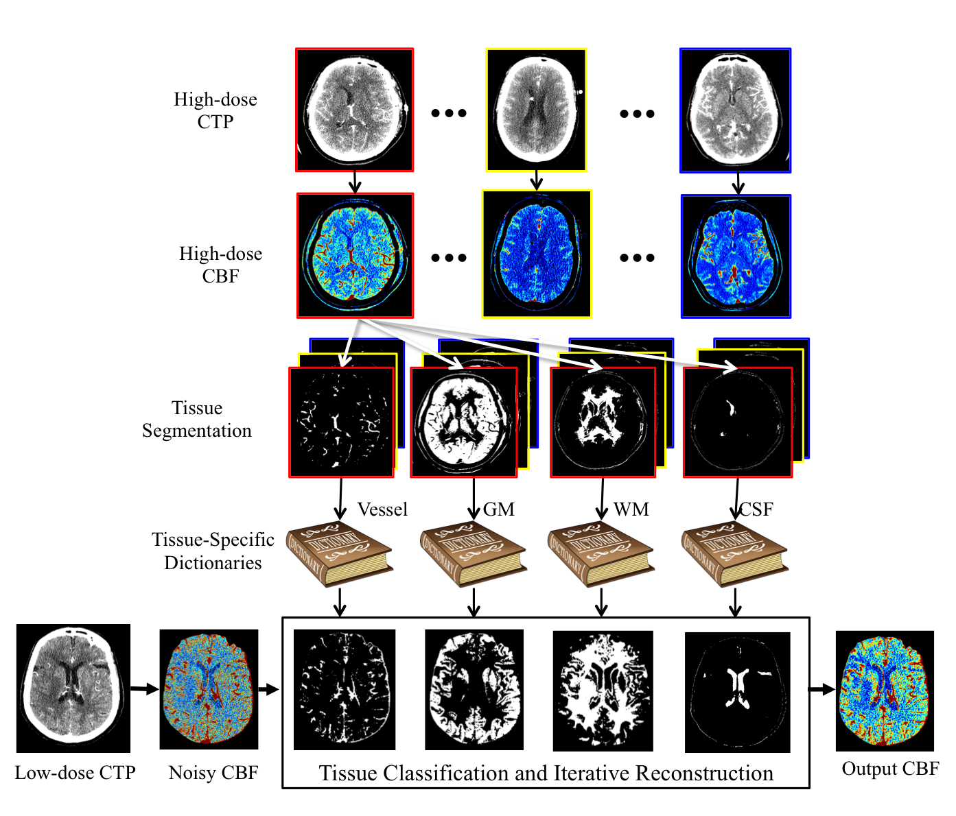
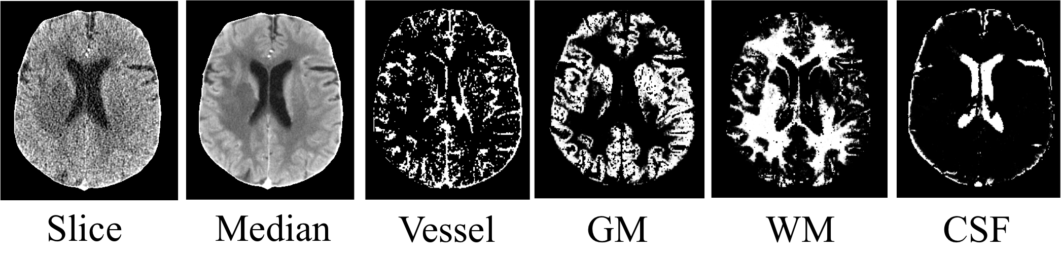
- STEP 1:Tissue Classification. Classify the voxels in CTP into four classes: vessel, gray matter (GM), white matter (WM) and cerebrospinal fluid (CSF).
Use Expectation-Maximization Segmentation (EMS) with contexture information incorporated by a MRF.
- STEP 2: Tissue-Specific Dictionaries Learning. Use online dictionary learning. Patches with 50% or more pixels in class i are used for training dictionary Di.
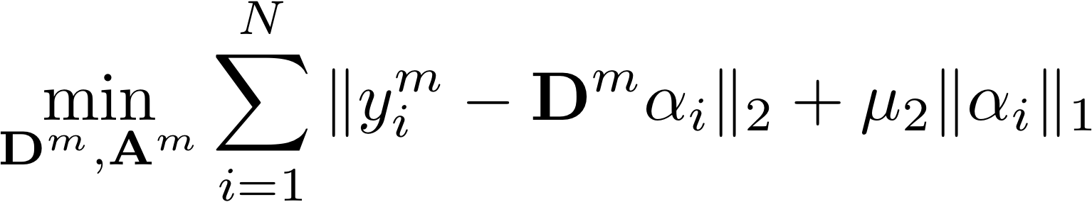
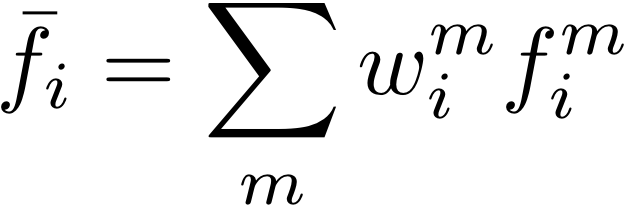
- STEP 3: Weighted Sparse Deconvolution. Reconstruct the CBF map for each tissue class using the respective dictionary.

Weighted sum for each pixel based on the classification probability map.
Results
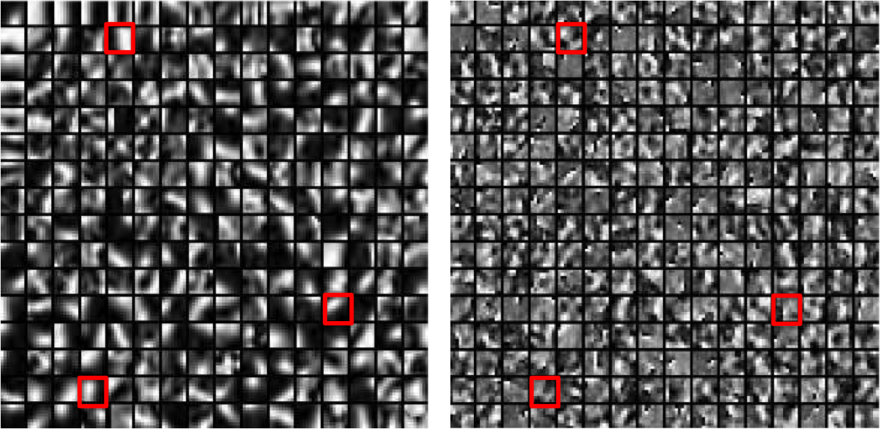
1. Learned dictionaries
Left is the global dictionary learned from all tissue classes. Right is the tissue-specific dictionary learned from white matter. The global dictionary is dominated by high-contrast, edge-like atoms, while the tissue-specific dictionary for WM has more low- contrast, fine structured atoms, as highlighted by red boxes.
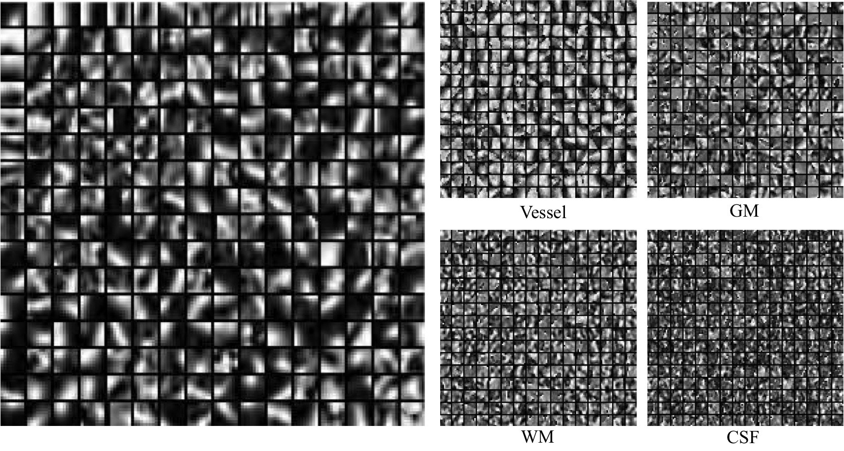
Here we show the global dictionary (left) and the four tissue-specific dictionaries (right).
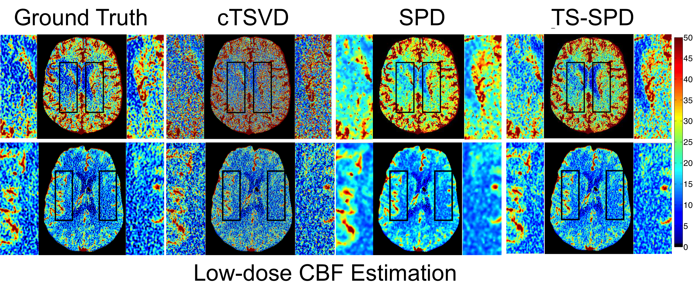
2. Visual Results of CBF Maps:
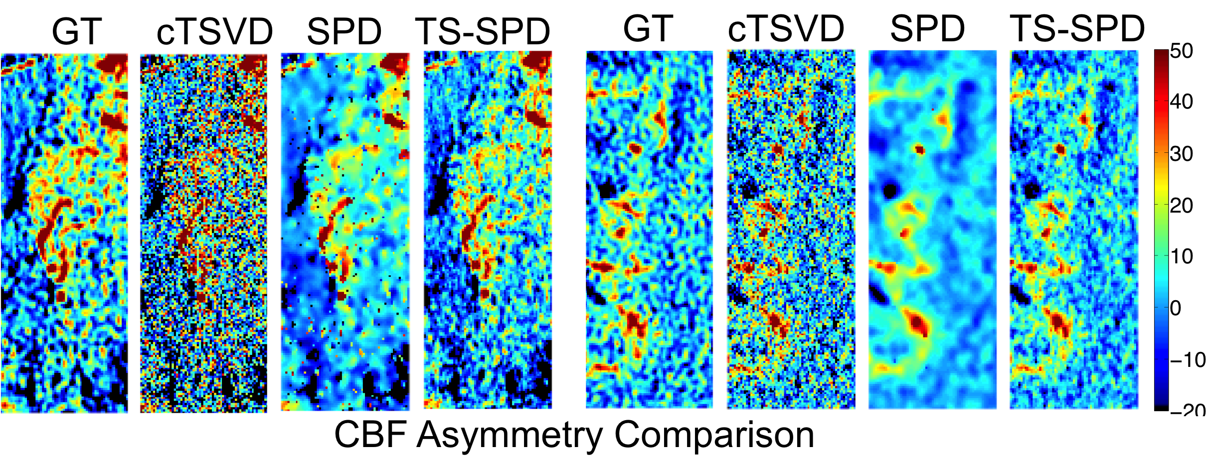
3. Asymmetry:
We compute the intensity difference maps between LMCA and RMCA for three methods.
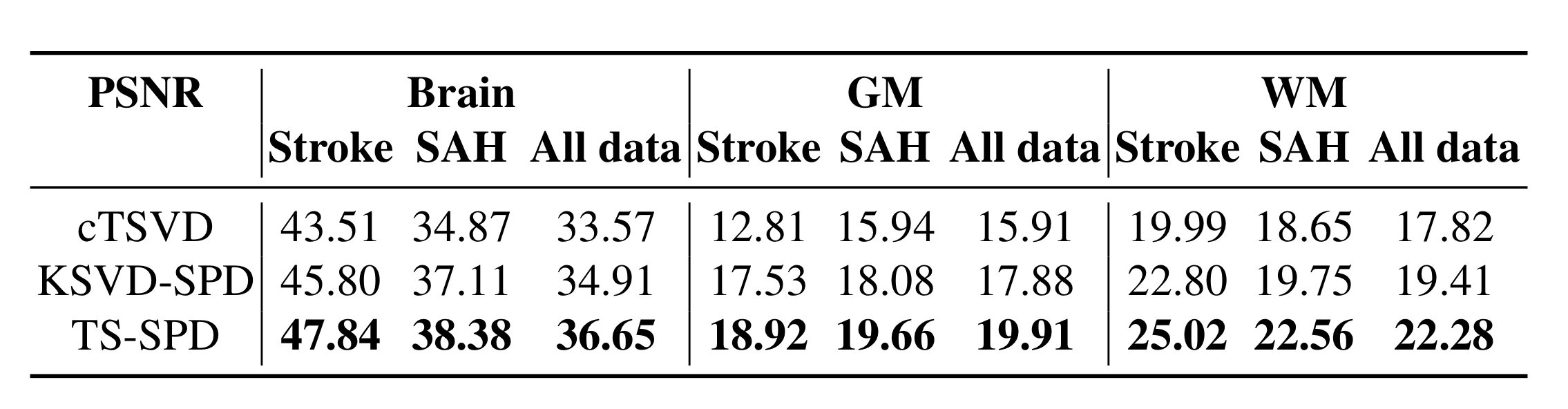
4. Quantitative Results:
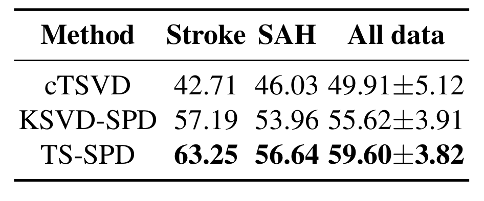
5. Ischemic/Normal voxels Separation:
Publication
- Ruogu Fang, Tsuhan Chen, Pina C. Sanelli. Tissue-Specific Sparse Deconvolution for Low-Dose CT Perfusion. Medical Image Computing and Computer-Assisted Intervention, MICCAI 2013. Lecture Notes in Computer Science.(MICCAI'13) [PDF]
Citation
- Ruogu Fang, Tsuhan Chen, Pina C. Sanelli. Tissue-Specific Sparse Deconvolution for Low-Dose CT Perfusion. Medical Image Computing and Computer-Assisted Intervention, MICCAI 2013. Lecture Notes in Computer Science.
Bibtex
@incollection{fang2012sparsity,
title={Tissue-Specific Sparse Deconvolution for Low-Dose CT Perfusion},
author={Fang, Ruogu and Chen, Tsuhan and Sanelli, Pina C},
booktitle={Medical Image Computing and Computer-Assisted Intervention--MICCAI 2012},
pages={272--280},
year={2012},
publisher={Springer}
}