Tissue-Specific Sparse Deconvolution for Low-Dose CT Perfusion MICCAI 2013
Abstract
Sparse perfusion deconvolution has been recently proposed to effectively improve the image quality and diagnostic accuracy of low-dose perfusion CT by extracting the complementary information from the high-dose perfusion maps to restore the low-dose using a joint spatio-temporal model. However the low-contrast tissue classes where infarct core and ischemic penumbra usually occur in cerebral perfusion CT tend to be over-smoothed, leading to loss of essential biomarkers. In this paper, we extend this line of work by introducing tissue-specific sparse deconvolution to preserve the subtle perfusion information in the low-contrast tissue classes by learning tissue-specific dictionaries for each tissue class, and restore the low-dose perfusion maps by joining the tissue segments reconstructed from the corresponding dictionaries. Extensive validation on clinical datasets of patients with cerebrovascular disease demonstrates the superior performance of our proposed method with the advantage of better differentiation between abnormal and normal tissue in these patients.
Downloads
BibTeX
@incollection{Fang_MICCAI12_Tissue,
   title = {Tissue-Specific Sparse Deconvolution for Low-Dose CT Perfusion},
  author = {Fang, Ruogu and Chen, Tsuhan and Sanelli, Pina C},
  booktitle = {Medical Image Computing and Computer-Assisted Intervention--MICCAI 2013},
  year = {2013},
  publisher = {Springer}
Publications on Robust CT Perfusion
Ruogu Fang, Tsuhan Chen, Pina Sanelli: Tissue-Specific Sparse Deconvolution for Low-Dose CT Perfusion. International Conference on Medical Image Computing and Computer Assisted Intervention. 2013. Lecture Notes in Computer Science.
Ruogu Fang, Tsuhan Chen, Pina Sanelli: Towards Robust Deconvolution of Low-Dose Perfusion CT: Sparse Perfusion Deconvolution Using Online Dictionary Learning. Medical Image Analysis 17(4): 417-428 (2013) (Top 25 hottest articles in Medical Image Analysis in 2013 April to June [Link] )
Ruogu Fang, Tsuhan Chen, Pina Sanelli: Sparsity-Based Deconvolution of Low-Dose Perfusion CT Using Learned Dictionaries. Medical Image Computing and Computer-Assisted Intervention – MICCAI 2012. Lecture Notes in Computer Science Volume 7510, 2012, pp 272-280 (MICCAI)
Ruogu Fang, Tsuhan Chen, Pina Sanelli. Sparsity-Based Deconvolution Of Low-Dose Brain Perfusion CT In Subarachnoid Hemorrhage Patients. In Proceeding of the 9th International Symposium on Biomedical Imaging, pp. 872-875, 2012. (ISBI) (Oral presentation)
Ruogu Fang, Ashish Raj, Tsuhan Chen, Pina C. Sanelli. Radiation dose reduction in computed tomography perfusion using spatial-temporal Bayesian methods. In Proceedings of SPIE Medical Imaging, Volume 8313, Paper #831345, 2012. (SPIE)
Motivation
Theoretically, a global dictionary is able to capture sufficient image information for different tissue classes, given abundant training data from each class. However empirically the optimization procedure which minimizes the overall reconstruction error, tends to favor high-contrast patches to the low-contrast ones in both the learning and reconstruction procedures. For medical images, the subtle variations and changes embedded in the low-contrast tissue classes such as white matter can be crucial for disease detection and diagnosis.Contributions
- Tissue-specific dictionaries for each tissue class are employed in place of the global dictionary to capture the low-contrast tissue class and delicate structural details.
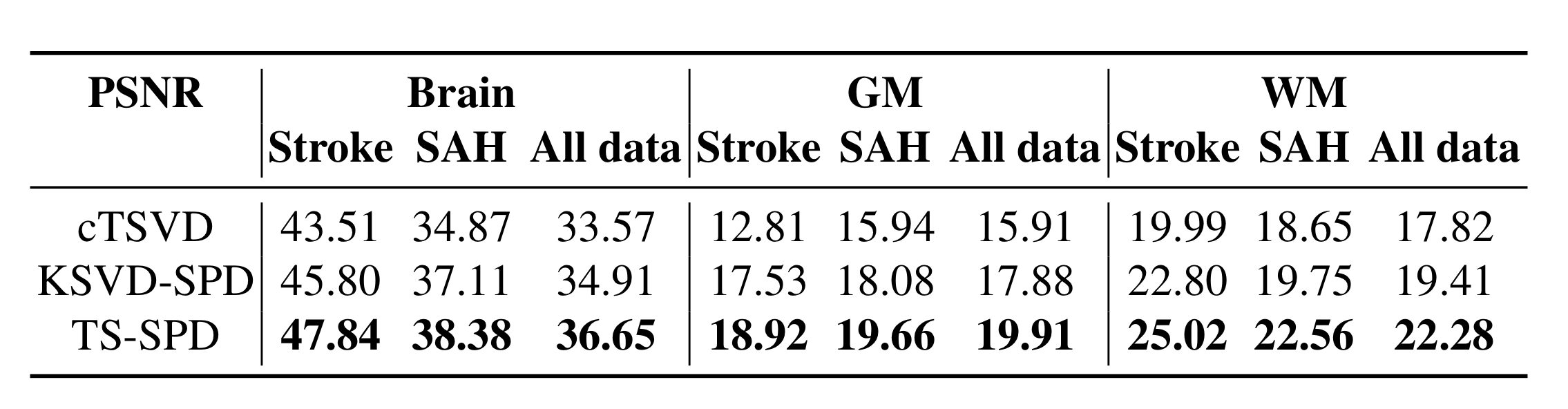
- Weighted sparse deconvolution based on the probability of the tissue classification is proposed for a unified reconstruction of the low-dose perfusion maps. In vivo brain acute stroke and aneurysmal SAH patients data, we demonstrate the superiority of our proposed method in CBF estimation that leads to better separation between normal and ischemic tissue.
Algorithm
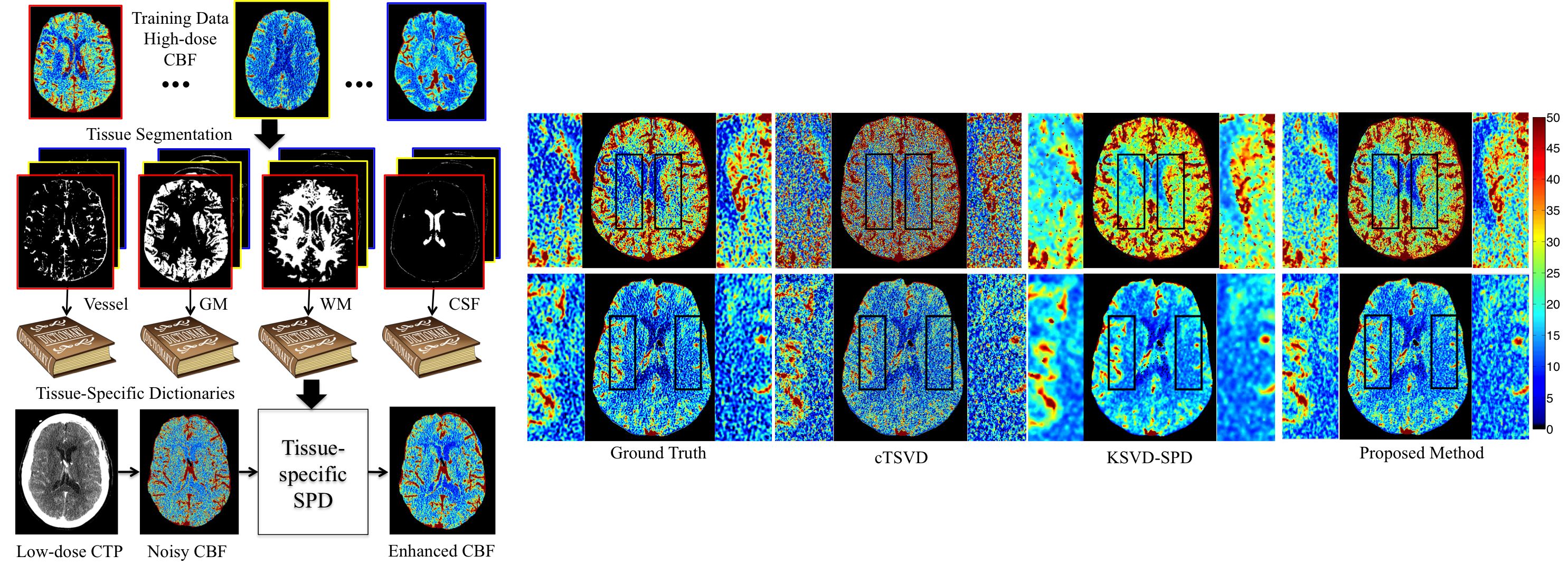
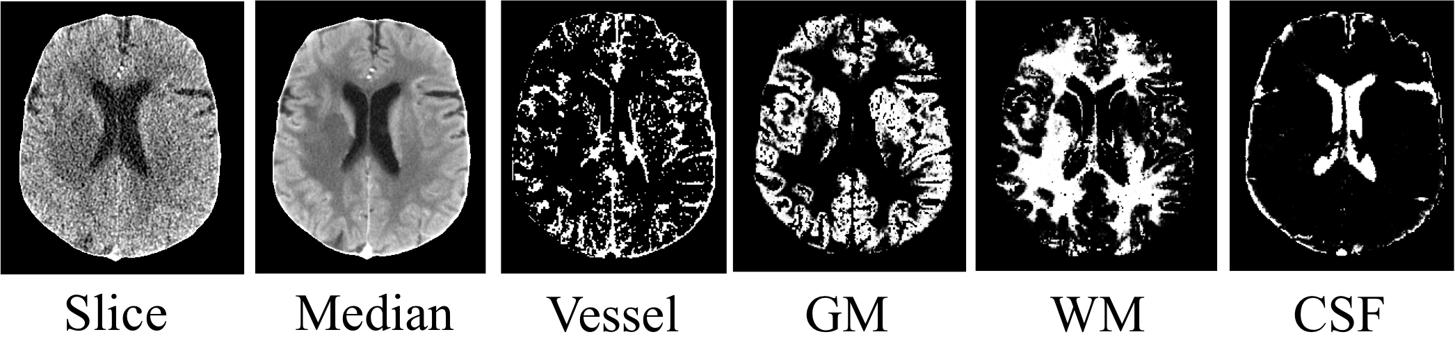
- STEP 1:Tissue Classification. Classify the voxels in CTP into four classes: vessel, gray matter (GM), white matter (WM) and cerebrospinal fluid (CSF).
Use Expectation-Maximization Segmentation (EMS) with contexture information incorporated by a MRF.
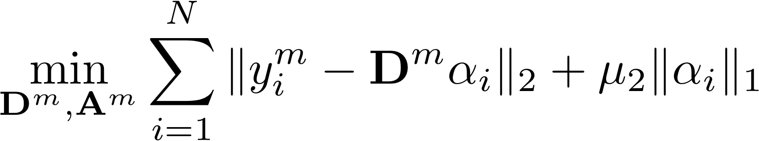
- STEP 2: Tissue-Specific Dictionaries Learning. Use online dictionary learning. Patches with 50% or more pixels in class i are used for training dictionary Di.
- STEP 3: Weighted Sparse Deconvolution. Reconstruct the CBF map for each tissue class using the respective dictionary.

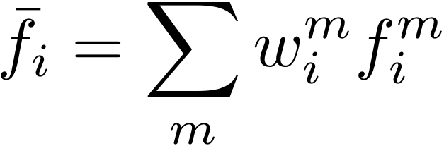
Weighted sum for each pixel based on the classification probability map.
Results
1. Learned dictionaries
Left is the global dictionary learned from all tissue classes. Right is the tissue-specific dictionary learned from white matter. The global dictionary is dominated by high-contrast, edge-like atoms, while the tissue-specific dictionary for WM has more low- contrast, fine structured atoms, as highlighted by red boxes.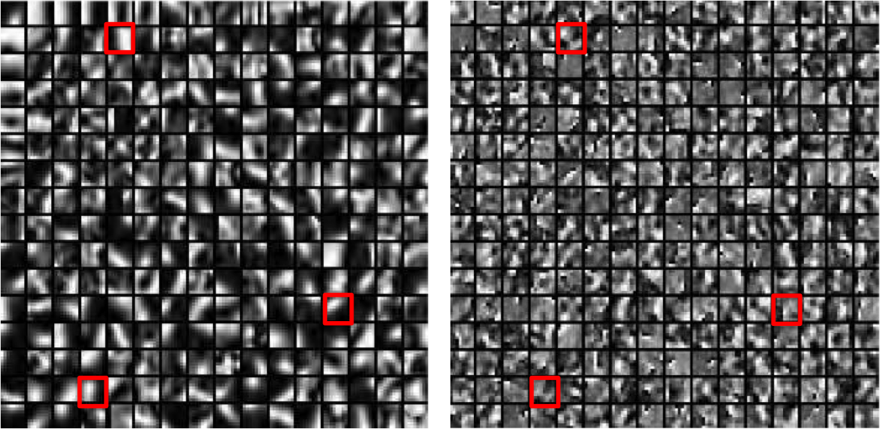
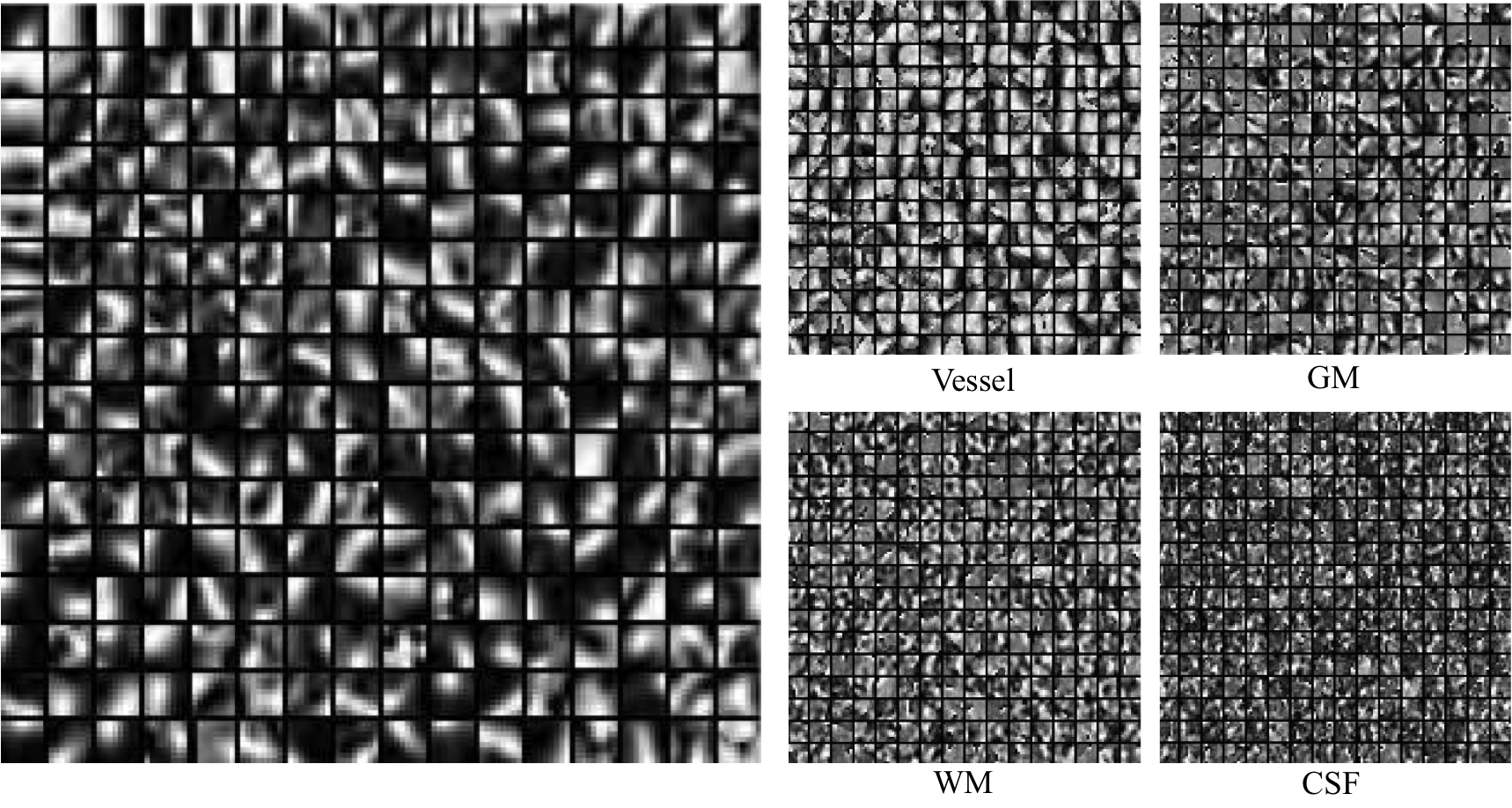
Here we show the global dictionary (left) and the four tissue-specific dictionaries (right).
2. Visual Results of CBF Maps:
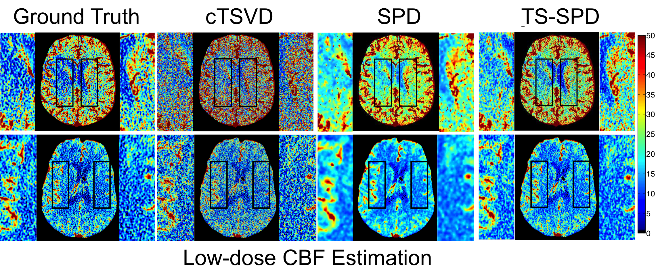
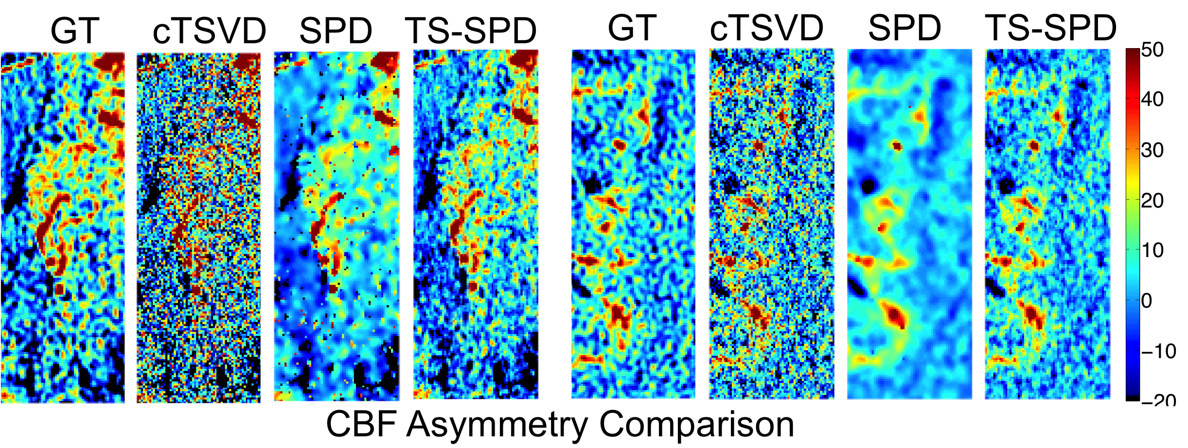
3. Asymmetry:
We compute the intensity difference maps between LMCA and RMCA for three methods.
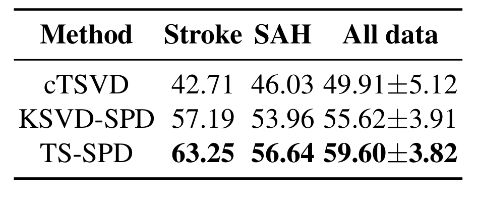
4. Quantitative Results:
5. Ischemic/Normal voxels Separation: